Figure 1
Download original image
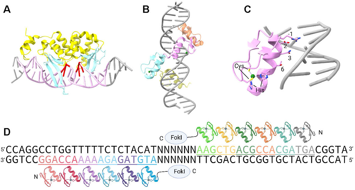
(A) The structure of I-SceI bound to the DNA substrate (PDB ID: 1R7M). The meganuclease is yellow, while its multiple β-strands interacting with DNA bases are colored cyan. The 18-bp DNA sequence recognized by I-SceI is in pink, and the DNA cleavage sites are in red. The cartoon is depicted with ChimeraX 1.3. (B) Tandem zinc-finger repeats with the target DNA (PDB ID: 2I13). (C) An individual zinc finger repeat interacting with DNA. Key protein residues responsible for DNA base recognition and zinc ion coordination are shown as sticks. The zinc ion is presented as a green sphere. (D) Schematic diagram of ZFN. ZF modules are indicated in different colors, and DNA triplets are underlined and shown in the same color.
Current usage metrics show cumulative count of Article Views (full-text article views including HTML views, PDF and ePub downloads, according to the available data) and Abstracts Views on Vision4Press platform.
Data correspond to usage on the plateform after 2015. The current usage metrics is available 48-96 hours after online publication and is updated daily on week days.
Initial download of the metrics may take a while.